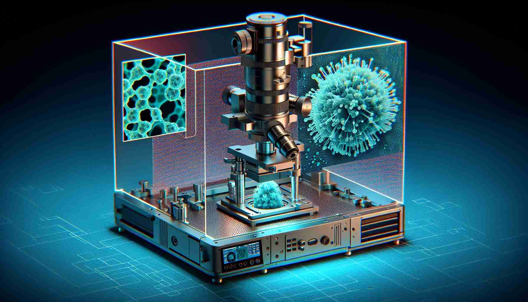
Researchers have made groundbreaking advancements in Atomic Force Microscopy (AFM) technology, paving the way for visualizing flexible 3D structures with high precision. By utilizing a cutting-edge extension of AFM, scientists have successfully imaged a suspended nanostructure in 3D, showcasing the immense potential of this technique in studying various biological systems.
Gone are the days of limited 2D imaging capabilities, as AFM now offers the ability to delve into the intricate details of living cells and molecular structures in a 3D space. Through innovative experiments and simulations, researchers have demonstrated that AFM can accurately capture the topography and interactions of nanosized objects, including carbon nanotube fibers and platinum nanodots.
The key lies in employing dynamic mode AFM, where a vibrating tip interacts with the sample’s surface, reducing the risk of damage and enabling precise imaging. This mode allows for a more detailed analysis of the forces at play, shedding light on the imaging mechanisms crucial for understanding complex biological systems.
With these recent developments, the realm of nanoscale imaging is undergoing a revolution, offering scientists a powerful tool to explore the depths of life phenomena. The future of AFM holds promise for unlocking new insights into cells, organelles, chromosomes, and vesicles, paving the way for transformative discoveries in the field of biology.
Revolutionizing AFM Technology for 3D Biological Imaging: Unveiling New Frontiers
In the realm of Atomic Force Microscopy (AFM) technology for 3D biological imaging, groundbreaking progress continues to shape the landscape of scientific exploration. While the previous article touched upon the advancements in imaging flexible 3D structures with high precision, there are additional critical aspects that deserve attention to provide a comprehensive view on this evolving field.
Key Question: How does AFM technology cope with imaging biological samples with varying stiffness and topographical complexity?
Answer: Traditional AFM techniques may face challenges when dealing with biological samples that exhibit different levels of stiffness and intricate topographical features. To address this, researchers are exploring novel approaches such as multifrequency AFM and high-speed AFM to enhance imaging capabilities across a broader range of biological specimens.
Key Challenge: One of the primary challenges associated with revolutionizing AFM technology for 3D biological imaging is the substantial data processing requirements for reconstructing intricate 3D structures from AFM scans.
Advantages:
– AFM technology provides unparalleled resolution and precision in capturing the 3D topography of biological samples at the nanoscale level.
– The non-destructive nature of AFM imaging enables repeated scans of living cells and delicate biological structures without causing harm.
– Dynamic mode AFM offers a versatile platform for studying dynamic processes within biological systems in a 3D context.
Disadvantages:
– AFM imaging can be time-consuming, particularly when scanning large areas or complex biological structures.
– High-resolution AFM imaging often requires a high level of expertise to optimize imaging parameters and data interpretation accurately.
– The cost associated with acquiring and maintaining advanced AFM systems may be prohibitive for some research laboratories.
As researchers push the boundaries of AFM technology for 3D biological imaging, collaborations between interdisciplinary teams and the integration of machine learning algorithms for data analysis are emerging as vital strategies to overcome existing challenges. The transformative potential of AFM in unraveling the mysteries of biological systems remains a driving force for continued innovation and exploration.
For more insights on cutting-edge AFM applications and developments, visit nanoscience.com.


